Structure in Fish, Amphibians, Birds, and Reptiles

Fishedit
The epidermis of fish and of most amphibians consists entirely of live cells, with only minimal quantities of keratin in the cells of the superficial layer. It is generally permeable, and in the case of many amphibians, may actually be a major respiratory organ. The dermis of bony fish typically contains relatively little of the connective tissue found in tetrapods. Instead, in most species, it is largely replaced by solid, protective bony scales. Apart from some particularly large dermal bones that form parts of the skull, these scales are lost in tetrapods, although many reptiles do have scales of a different kind, as do pangolins. Cartilaginous fish have numerous tooth-like denticles embedded in their skin, in place of true scales.
Sweat glands and sebaceous glands are both unique to mammals, but other types of skin gland are found in other vertebrates. Fish typically have a numerous individual mucus-secreting skin cells that aid in insulation and protection, but may also have poison glands, photophores, or cells that produce a more watery, serous fluid. In amphibians, the mucus cells are gathered together to form sac-like glands. Most living amphibians also possess granular glands in the skin, that secrete irritating or toxic compounds.
Although melanin is found in the skin of many species, in the reptiles, the amphibians, and fish, the epidermis is often relatively colorless. Instead, the color of the skin is largely due to chromatophores in the dermis, which, in addition to melanin, may contain guanine or carotenoid pigments. Many species, such as chameleons and flounders may be able to change the color of their skin by adjusting the relative size of their chromatophores.
Amphibiansedit
See also: amphibians
Overviewedit
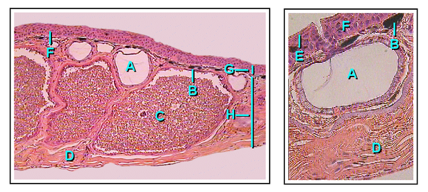
Amphibians possess two types of glands, mucous and granular (serous). Both of these glands are part of the integument and thus considered cutaneous. Mucous and granular glands are both divided into three different sections which all connect to structure the gland as a whole. The three individual parts of the gland are the duct, the intercalary region, and lastly the alveolar gland (sac). Structurally, the duct is derived via keratinocytes and passes through to the surface of the epidermal or outer skin layer thus allowing external secretions of the body. The gland alveolus is a sac shaped structure that is found on the bottom or base region of the granular gland. The cells in this sac specialize in secretion. Between the alveolar gland and the duct is the intercalary system which can be summed up as a transitional region connecting the duct to the grand alveolar beneath the epidermal skin layer. In general, granular glands are larger in size than the mucous glands, however mucous glands hold a much greater majority in overall number.
Granular Glandsedit
Granular glands can be identified as venomous and often differ in the type of toxin as well as the concentrations of secretions across various orders and species within the amphibians. They are located in clusters differing in concentration depending on amphibian taxa. The toxins can be fatal to most vertebrates or have no effect against others. These glands are alveolar meaning they structurally have little sacs in which venom is produced and held before it is secreted upon defensive behaviors.
Structurally, the ducts of the granular gland initially maintain a cylindrical shape. However, when the ducts become mature and full of fluid, the base of the ducts become swollen due to the pressure from the inside. This causes the epidermal layer to form a pit like opening on the surface of the duct in which the inner fluid will be secreted in an upwards fashion.
The intercalary region of granular glands is more developed and mature in comparison with mucous glands. This region resides as a ring of cells surrounding the basal portion of the duct which are argued to have an ectodermal muscular nature due to their influence over the lumen (space inside the tube) of the duct with dilation and constriction functions during secretions. The cells are found radially around the duct and provide a distinct attachment site for muscle fibers around the gland's body.
The gland alveolus is a sac that is divided into three specific regions/layers. The outer layer or tunica fibrosa is composed of densely packed connective-tissue which connects with fibers from the spongy intermediate layer where elastic fibers, as well as nerves, reside. The nerves send signals to the muscles as well as the epithelial layers. Lastly, the epithelium or tunica propria encloses the gland.
Mucous Glandsedit
Mucous glands are non-venomous and offer a different functionality for amphibians than granular. Mucous glands cover the entire surface area of the amphibian body and specialize in keeping the body lubricated. There are many other functions of the mucous glands such as controlling the pH, thermoregulation, adhesive properties to the environment, anti-predator behaviors (slimy to the grasp), chemical communication, even anti-bacterial/viral properties for protection against pathogens.
The ducts of the mucous gland appear as cylindrical vertical tubes that break through the epidermal layer to the surface of the skin. The cells lining the inside of the ducts are oriented with their longitudinal axis forming 90-degree angles surrounding the duct in a helical fashion.
Intercalary cells react identically to those of granular glands but on a smaller scale. Among the amphibians, there are taxa which contain a modified intercalary region (depending on the function of the glands), yet the majority share the same structure.
The alveolor of mucous glands are much more simple and only consist of an epithelium layer as well as connective tissue which forms a cover over the gland. This gland lacks a tunica propria and appears to have delicate and intricate fibers which pass over the gland's muscle and epithelial layers.
Birds and reptilesedit
The epidermis of birds and reptiles is closer to that of mammals, with a layer of dead keratin-filled cells at the surface, to help reduce water loss. A similar pattern is also seen in some of the more terrestrial amphibians such as toads. However, in all of these animals there is no clear differentiation of the epidermis into distinct layers, as occurs in humans, with the change in cell type being relatively gradual. The mammalian epidermis always possesses at least a stratum germinativum and stratum corneum, but the other intermediate layers found in humans are not always distinguishable. Hair is a distinctive feature of mammalian skin, while feathers are (at least among living species) similarly unique to birds.
Birds and reptiles have relatively few skin glands, although there may be a few structures for specific purposes, such as pheromone-secreting cells in some reptiles, or the uropygial gland of most birds.
Comments
Post a Comment